Skin pigmentation studies for cosmetics
Skin pigmentation is still a biological mechanism of interest in the cosmetic field. Whether it is to lighten the skin, provide a tanning effect or homogenize the complexion, brands and ingredient suppliers are constantly looking for ever more active products. Research on the mechanisms involved in skin pigmentation has made it possible to identify new pathways that are just as much targets for new active cosmetic ingredients.
We have set up models to carry out solid studies on skin pigmentation to validate the product’s effect and identify its mechanism of action.

Skin melanocytes synthetize a pigment, the melanin, which is transferred via melanocytes dendrites to keratinocytes. Melanin transfer results in skin pigmentation darkening of the skin, but also creating age spots.
Although melanin plays an important role in photoprotection against UV-induced DNA damages, melanin production can be altered by external stresses (sun exposure, pollution, …) and internal factors (hormones, inflammation, aging). Such malfunction can lead to pigmentation defect that has to be treated with adequate skin care products. Improvement of the skin color homogeneity can be provided by cosmetic products and need to be demonstrated by solid research studies.
Syntivia as an expert company in the research and development of cosmetic active ingredients is developping various models to study both activation and down regulation of skin pigmentation.
Skin models
Primary cells in culture
Proper conditions allow the cells in culture to live, grow and maintain their functions as if they were within the skin.
Syntivia uses primary cells, normal human melanocytes in 2D culture and in co-culture with normal human keratinocytes. Cells are isolated from skin of different phototypes to reproduce the human skin configuration.
Our equipment allows us to obtain reliable results on this model: gene/protein expression, melanin quantification, melanin transfer and melanocyte dendricity.
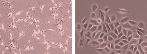
Normal Human Epidermal Melanocytes and Normal Human Epidermal Keratinocytes in 2D culture.
Epidermal microtissues
The epidermal microtissue is a mixed cellular model composed of the two main cell types of the epidermis: melanocytes and keratinocytes. This model is created from human primary cells grown as spheroids; we can choose the age and phototype of the donor. This model is produced and cultivated in 96-well plates, making it suitable for screening. This is an epidermis model that can be adapted to specific issues: pigmentation, inflammation, toxicity.
Epidermis microtissue composed of NHEM and NHEK.
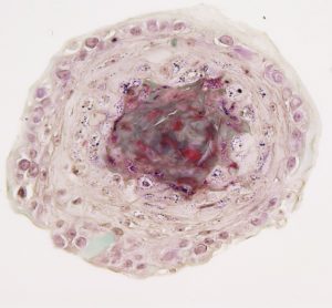
Skin explants
Syntivia uses a skin explant model obtained by plastic surgery. The skin explant is kept alive for 7 days to allow ex vivo pigmentation studies to predict in vivo skin response.
Skin explant
Skin pigmentation studies
Study of pigmentation biological pathways
We use genomic chips on genes involved in each of the pigmentation processes. We can work on all our skin models. We use the Fluidigm’s BiomarkTM technology, this high-tech tool based on microfluidic technology allows to obtain:
- A large amount of data
- Flexible conditions
- Precise quantification of gene expression
The system’s technoloy is robust and yields reliable statistics on a wide range of targeted genes.
Cosmetogenomic chips have been especially designed to test active ingredients or cosmetic formulas.
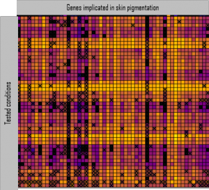
Cosmetogenomic chip results: activation and inhibition of gene expression on skin explant.
Melanin synthesis: induction and inhibition of melanin production
We are able to reliably quantify melanin synthesis in human skin cells. It allows to identify compounds capable of activating or inhibiting melanin neosynthesis.
Models used: primary cells and epidermis microtissue.
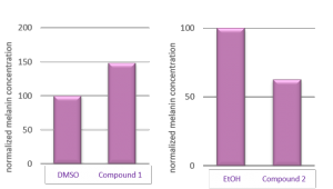
Melanin content assay. Melanin production can be quantified after treatment with active compounds by spectroscopy.
Melanin transfer: Imaging and quantification
Melanin is transmitted from melanocytes to keratinocytes by cellular vesicles called melanosomes. Using fluorescence microscopy and flow cytometry, we can visualize and quantify the transfer of melanosomes from one cell type to another in 2D or 3D models.
Models used: primary cells in co-culture and epidermis microtissue.
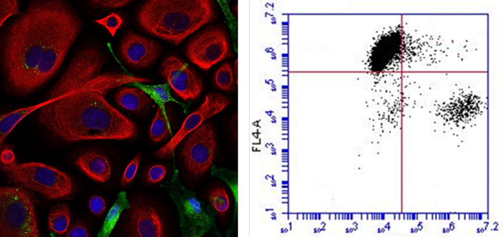
Left: Melanosome transfer from melanocytes to keratinocytes visualized by fluorescent microscope in 2D after specific labelling (co-culture of melanocytes and keratinocytes). Right: Melanin transfer from melanocytes to keratinocytes can be reliably quantified by flow cytometry after specific labelling (dissociated microtissue).
Melanocyte dendricity: Imaging and quantification
Melanin transfer is directly connected to melanocyte dendricity. The melanosomes containing melanin are transported by a network of microtubules to the dendritic ends of melanocytes. In order to ensure the transfer of melanosomes to the cytoplasm of keratinocytes, melanocytes dendrites creep in between neighbouring keratinocytes. We can visualize and quantify melanocyte dendricity.
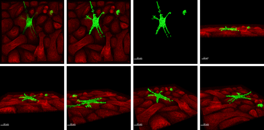
Co-culture of melanocytes and keratinocytes activated for melanin transfer, acquired with a confocal microscope. The Imaris software was used to proceed to the surfacing of melanocytes and 3D imaging.
The cell morphology can be visualized in 3D on epidermis microtissue by SPIM (Selective Plan Illumination Microscopy). The cells are labelled prior to spheroid formation with melanocytes in green and keratinocytes in red.
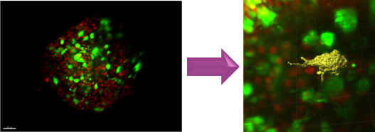
Observation of melanocyte dendricity in epidermal spheroid by 3D surfacing.
Pigmentation studies directly on human skin
Proteins implicated in melanin synthesis can be quantified by fluorescent labelling in skin explants and linked with melanin production.
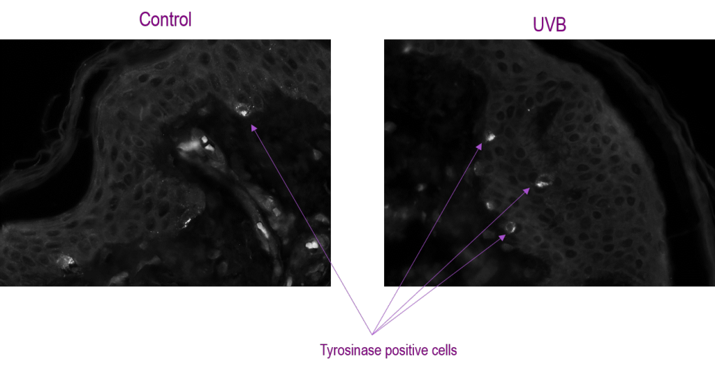
Labelling of Tyrosinase in a skin section observed by fluorescent microscope.
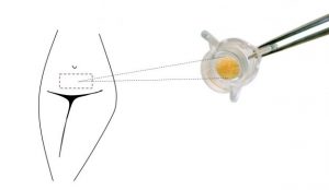
Skin explants can be treated topically with any cosmetic compound. Melanin production is observed by histology and quantified by image processing.
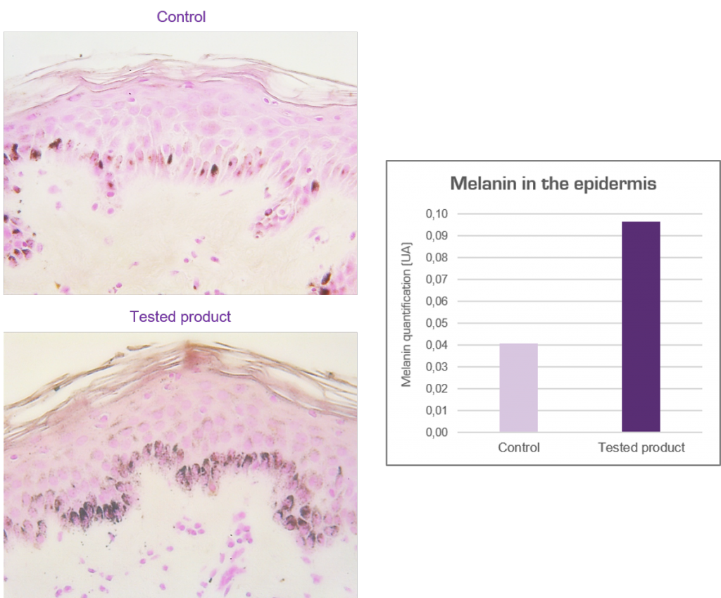
Treatment with test product twice a day for 7 days and quantification of melanin in the skin after coloration.
Our expertise in skin pigmentation study has been built through the development of many active ingredients and cosmetic products. Do not hesitate to share your projects with us so that we can set up a study adapted to your needs.
The Syntivia team is completing these studies with more details, do not hesitate to contact us for more information on pigmentation studies.