New 3D skin model for cosmetic in vitro tests
The epidermal spheroid, a new 3D skin model
As the cosmetic sector no longer allows the use of animal experiments to validate the effect of compounds, finding intermediate models between monolayer cell cultures and live human organisms has become a central issue. Marine Norlund, Biology Project Manager at Syntivia, has presented her work on a new 3D model for preclinical toxicological studies at the Centre Pierre Poitier in Toulouse at 11am on May 20th, 2016,
A new alternative to animal testing
The need to find innovative models to study toxicological effects prior to clinical trials has become particularly urgent for the cosmetic industry. Today, the only OECD approved alternative to animal tests for toxicology is reconstructed epidermis. In this context, Syntivia conducted research on a new 3D model of the human epidermis, in collaboration with the ITAV Institute for Advanced Life Science and Toulouse Tech Transfer.
The project resulted in the creation and validation of a new and completely characterized epidermis model that is perfectly functional for in-vitro studies. The unique 3D skin model is composed of keratinocytes and melanocytes, the two major cell types found in the epidermis. The epidermal spheroid is closer to the microenvironment of human skin than 2D monolayer cultures and less complicated in use than reconstructed skin and human skin biopsies.
Faithful reproduction of the epidermis microenvironment
In the first days of culture, the epidermal spheroid is a very good model to study melanogenesis. We have developed a method to evaluate the total amount of melanin within the epidermal spheroid and are able to modulate the melanin production by reference treatments. Using 3D microscopy, melanin transfer from melanocytes to keratinocytes can be visualized within the spheroid.
Within time in culture, the epidermal spheroid becomes a well-organized organelle with physiological keratinocyte differentiation. After 7 days of culture, the epidermal spheroid faithfully reproduces the organization and functionality of the human epidermis. The proliferated cells are specifically located at the periphery of the spheroid as in the basal layer and the keratinocyte differentiation reproduce the corneal layer to the inside of the spheroid (figure 1).
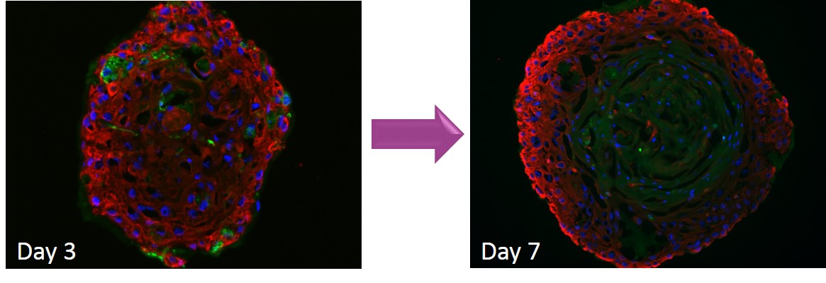 Figure 1: Organization of the spheroid in an epidermis-like organelle in 7 days of culture
The epidermal spheroid is functional for the analysis of the melanogenesis pathway and differentiation and allows us to propose this model for the development or study of active molecules for cosmetic or pharmacologic applications. The epidermal spheroid is a faithful epidermis equivalent (figure 2) and a good alternative to reconstructed epidermis: it’s easier to produce, less expensive and gives full physiological response.
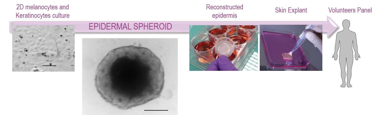 Figure 2: Positioning of the epidermal spheroid among skin models