High-resolution Skin imaging
High resolution skin imaging to illustrate cosmetic claims
The combination of biphoton and confocal microscopy opens up a wide range of possibilities for beautiful in-depth skin imaging to support cosmetic claims on any type of sample. The Syntivia team helps you obtain high quality 2D and 3D images to illustrate your ingredient’s properties with outstanding scientific examples. Here’s how it works:
Combining biphoton and confocal microscopy to increase precision
Classic microscopy doesn’t allow segmentation and has a limited level of precision. Confocal microscopy uses a classic laser to illuminate an entire sample. A “pinhole” diaphragm then enables to recover with high precision the light from a specific point (x;y;z), in the sample in order to split it up in small, segmented units.
Biphoton microscopes do the opposite. They only illuminate a specific point (x;y;z) in the sample and then collect the light emission from the entire sample. Biphoton microscopes are extremely powerful: they allow to study a sample in depth (up to 1 cm). As the technique only requires to illuminate one specific point, phototoxicity is very low. Fluorescent labelling can be detected at any wavelength.
Syntivia combines both techniques by using a biphoton/confocal microscope, a state-of-the-art device for hi-tech sample observation and in-depth imaging. The device is equipped with 2 optic heads: one to illuminate samples from the top, the other from the bottom. The experts at Syntivia use the biphoton/confocal technique to test active ingredients and cosmetic formulas and visualize their effects. 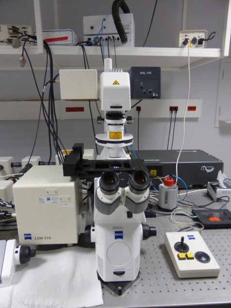 The biphoton confocal microscope used by Syntivia to perform high resolution skin imaging.
High resolution 2D and 3D images for any type of sample
Combined biphoton/confocal microscopy allows in-depth sample imaging and provides high-quality images to support cosmetic claims with outstanding examples. The 2D and 3D images illustrate your ingredient’s properties thanks to fluorescent labelling. As the biphoton/confocal microscope allows high contrast optical sections, it allows to detect fluorescent labelling on a wide range of models: living or fixed cells, epidermal microtissue or cleared skin explants. The technology offers a wide range of benefits:
- In-depth sample analysis
- Reliable quantification of fluorescent markers
- High quality 2D and 3D images thanks to Z sectioning
Biphoton/confocal microscope allows observing the effects of cosmetic ingredients on any type of sample, using 2 different microscopic techniques. Biphoton/confocal microscopes allow to obtain very precise in-depth images from both large samples, such as skin explants, spheroids as well as small samples, such as cells. Below you’ll find some illustrations of images the technology retrieves:
Analysis of skin cells in 2D and 3D
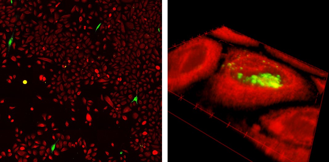
On the left: cell analysis in a co-culture of primary normal human epidermal keratinocytes (in red) and normal human melanocytes (in green). On the right: image obtained by confocal microscopy and 3D reconstruction of melanin (in green) transfer to keratinocytes (in red).
Biphoton confocal microscopy allows sample reconstruction. After optical sectioning, we assemble the images to recreate the 3D structure of the skin sample observed and provide our clients with beautiful images to support their cosmetic claims.
From 2D tissue sections to 3D reconstructed skin
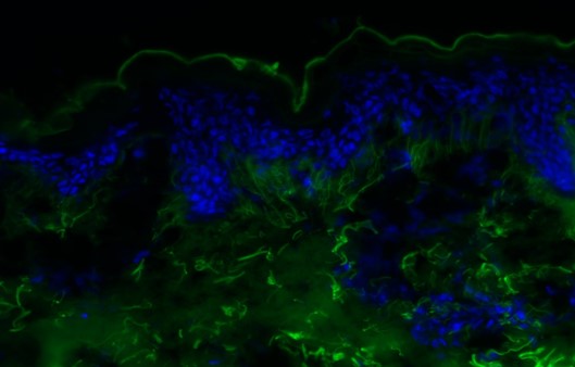 Autofluorescence of dermis and stratum corneum in skin explant measured by biphoton microscopy: optical section of skin (DAPI-labelled cell nuclei in blue, skin structure in green).
This precise optical section is followed by many others to detect changes in the different skin compartments. It is even possible to assemble the optical sections to obtain a 3D sample reconstruction and observe changes in the entire study model.
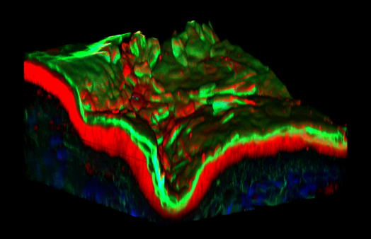 3D modeling skin explant: Collagen in blue (autofluorescence), epidermis in red (CMTMR labelling), stratum corneum and Elastin in green (autofluorescence).
The biphoton/confocal microscope provides detailed images using deep acquisition and provides high precision labelling from cell to tissue: all samples used can be labeled with specific or auto-fluorescence markers.
For more information on biphoton confocal microscopy and how we can use it to reveal your ingredient’s potential, don’t hesitate to contact us.