Human skin model to evaluate effect on premature aging
Syntivia recently published an article in the International Journal of Cosmetic Science, dealing with the development of one of its human skin models. This work, carried out in collaboration with the French National Centre for Scientific Research (CNRS) and the Toulouse Imaging Platform (TRI-Genotoul), has made it possible to develop a new study model that enables us to test not only products capable of protecting the skin from UV rays but also anti-aging products.

Human skin model of accelerated aging
UV rays are the main cause of premature skin aging. UV radiations cause DNA damage, apoptosis and deleterious morphological changes to the skin characteristic of photoaging.
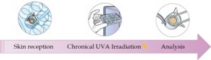
Experimental protocol to develop UVA exposed human skin model.
Human skin explants were chronically irradiated with UVA in order to mimic daily sunlight exposure. We used UVA for their ability to penetrate the skin deeply. Afterwards, effects on the epidermis were observed and quantified after histological staining and immunoflurescence. Skin morphology, as well as several markers involved in DNA repair and epidermal differentiation, were studied. Dermis deterioration was evaluated by transmission electronic microscopy and zymography in situ.
Epidermal disruption
The integrity of the epidermis is clearly affected by the UV treatment.
A disorganization of the layer is observed, characterized by a modification of cell cohesion and superposition. Moreover, the thickness of the epidermis decreased. The number of sunburn cells, that carry chromatin condensation within the nuclei, significantly increases.
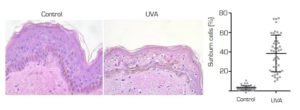
Morphological modifications of skin after UVA irradiation and sunburn cells quantification
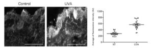
The histone H2AX becomes phosphorylated (ɣ-H2AX) in response to the formation of DNA breaks. On the irradiated skin, we detected a significant increase in positive ɣ-H2Ax stained nuclei representative of DNA damage.
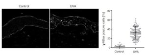
DNA damage observed and quantified after ɣ-H2AX labelling
The alteration of the intermediate keratinocyte differentiation state as well as the appearance of apoptotic cells confirm the deleterious and pro-aging effect on the epidermis.
Dermis disorganization
UV exposition causes a desassembly of the dermal fibers and a disruption of their organization.
The papillary and reticular dermis is formally altered by the treatment. The damage is characterized by a fragmentation of the fibers and a decrease in their density. Observed by transmission electronic microscopy, fibers are dispersed and noncohesive.
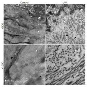
Extracellular matrix strucure observed by Transmission Electronic Microscopy (TEM)
Activation of proteases is responsible for the alteration of dermal fibers. UVA rays penetrating into the dermis are involved in this process of activation.
In irradiated skin, we have detected a strong increase in protease activity in both the epidermis and dermis. It was detected by in situ zymography.
In situ zymography of global skin proteases carried out on a cryosection of human skin explants
Evaluation of protective or repairing effects
Our human skin model is representative of premature aging and photoaging, characterized by epidermis alteration and accumulation of dermis malfunctions.
We have shown a strong alteration of the epidermis. The cells of the upper layers lose their cohesion, DNA lesions appear while keratinocyte differentiation is affected. We also observe disorganized dermis through the elastosis and the degradation of collagen I by matrix metalloproteases.
The life span of the skin explant is between 7 and 10 days. It allows skin markers to be measured only in the short term. Tested ingredient or product can be applied before, during or after UV exposition. The method of application chosen makes it possible to support a protective and/or repairing effect.
Don’t hesitate to contact us for more information on our other human skin models and product testing. You can share your projects with us so that we can set up a study adapted to your needs.