Cell screening
Large amounts of highly reliable data by cell screening
Our technology allows deep in cell imaging for protein and functional validation
Syntivia uses live cell screening to test active ingredients on a wide range of markers and activities. Our technology of choice is the ArrayscanTM automated microscope which allows us to run 90 test conditions in parallel during the same test.
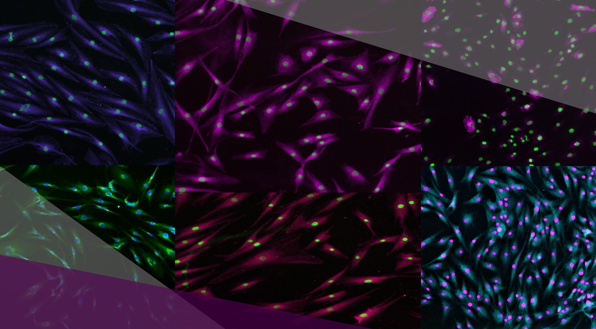
Images obtained by Arrayscan microscopy. This technology provides impressive images and very precise data.
This microscope technology is used to validate biological mechanisms induced by cosmetic ingredient(s) using functional screening. There are 2 ways to use it in a study :
– After a cosmetogenomic chip (genomic validation) for proteomic confirmation of the activity.
– Directly, with no previous step if you already have a good idea of your product’s properties and know what you want to demonstrate with high definition images and reliable data.
By trusting Syntivia to run cell screening on your compounds, you make the choice of excellence. Our highly qualified experts are professionals of the techniques we offer, and will provide you with precise results to substantiate your cosmetic claims or to refine your ingredient’s properties. We work in total objectivity to produce reliable reports and advise our clients on a biology and marketing view on their way to cosmetic development.
Our technology allows to quantify several types of markers : proteins, free radicals, DNA, cell metabolites (mitochondria, lysosomes)… If the targeted marker can be visualized via fluorescence or transmitted light, we will be able to quantify it and consequently target all types of activities:
– Barrier function (differentiation, adhesion, lipid production)
– Pigmentation
– Hair growth
– Elasticity
– Firmness
– Antimicrobial properties
– Moisturization
– Soothing properties
– Anti-oxidative defenses
– Energetic metabolism
– Extra-cellular matrix protection
– Detoxication
– Longevity
- This high tech automated microscope releases a large amount of highly reliable data on any cell markers.
- The statistics and 2D images obtained are highly reliable and will help you to illustrate your scientific communication
- The Arrayscan™ automated microscope yields reliable results on 96 conditions in parallel and limits human intervention, ensuring the total objectivity of the measurements obtained.
- It enables the visualization of biomarkers or changes in morphology on fixed or living cells.
The Arrayscan™ allows us to quantify any type of cell biomarker. This process helps us illustrate cytoplasm or nucleus protein, compartmental expression and cell morphology for cosmetic ingredient screening.
Syntivia works on primary skin cells obtained from all types of donors and kept in a 2D culture or co-culture.

Syntivia provides its clients with highly reliable results due to the quality of the equipment we use and the expertise of our collaborators. Arrayscan™ is an example of high technology for cell screening. The results are precise and accurately show the effect of cosmetic ingredients tested on live or fixed cells. Here are a few examples of results obtained with the Arrayscan™ technology.
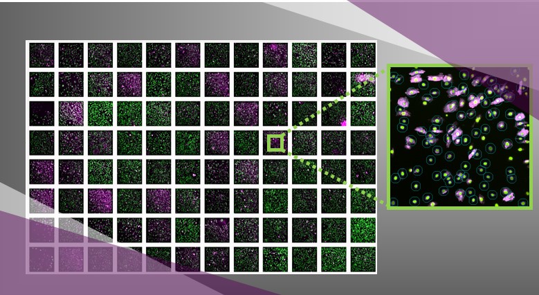
Quantification of Collagen I expression in 96-well plate by automated microscope Arrayscan™. Each well contains cells treated with a tested product. Several hundreds of cells can be analyzed by well.Cell nuclei are labelled in green, Collagen I in pink.The circles indicate the area where the fluorescent labelling is quantified (zoom on a field of a well).
With this analysis, it is possible to screen cosmetic ingredients on cell and visualize precisely if they have an effect thanks to the fluorescent labelling. We can see here that each tested product has a different effect on the cells. It is then possible to determine which cosmetic ingredient has the most interesting action and to go further with the study of this molecule of interest.
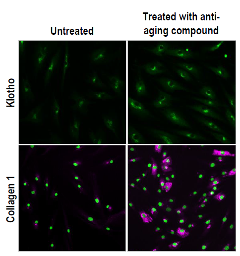
Quantification of 2 anti-aging proteins by automated microscope after treatment with an active molecule: Klotho in the endoplasmic reticulum (green) and Collagen I in the cytoplasm (pink).
In this example, the Arrayscan™ technology allows us to observe the effect of the tested compounds on the expression of Klotho and Collagen I proteins, which are both interesting targets in cosmetic ingredient research. This image has a double role : first, it clearly shows the effect of a cosmetic ingredient on protein expression. Second, these are beautiful images, perfectly usable in marketing documents to illustrate accurately and aesthetically the action of a product on skin cells. To support and make your documentation even more convincing, we can produce graphs using the Arrayscan™ numerous data to confirm your results with statistically verified numbers.
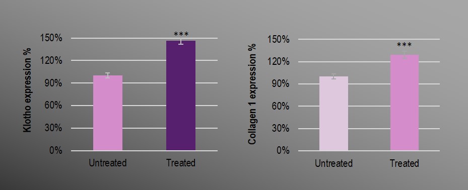
Graphic illustration of the protein expression quantification in human fibroblasts by Arrayscan™ microscopy.
The test conditions are totally customizable. The Arrayscan™ technology is a very useful tool for live cell screening and claim substantiation.
Contact us